This technique is used to visualise nanomaterials, and provides information on size and size distribution, shape, homogeneity and more.
Measurement principle
A beam of electrons is transmitted through a specimen (e.g. a suspension of particles on a grid for particle sizing, or an ultrathin section less than 100 nm thick for determining particle localisation in tissue). An image is formed from the interaction of the electrons with the sample as the beam is transmitted through the specimen.
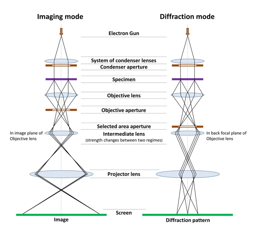
Figure: Schematic view of imaging and diffraction modes in TEM.
Image credit: Black Tubus
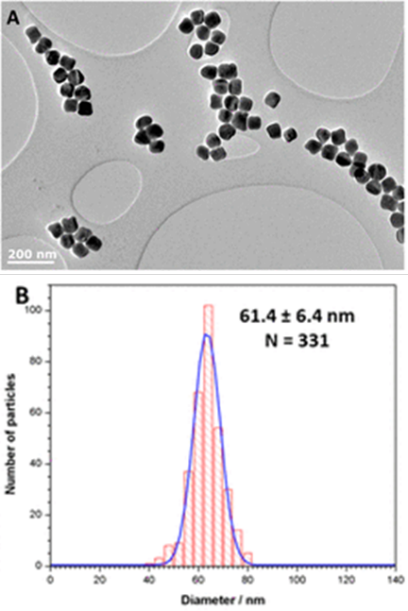
Image credit: Xianjin Cui, University of Birmingham.
Adapted from: https://doi.org/10.1021/acsami.9b03062
|
Particle characterisation (size, shape, homogeneity etc.)
- NMs are dispersed in ethanol or equivalent & deposited on TEM grid (e.g. holey carbon), air dried and imaged.
- For size / polydispersity at least 200 particles must be measured (with their diameter measured using software such as ImageJ) for statistical significance. Shape dimensions can also be determined.
- Note: It may be necessary to distinguish primary particles from agglomerates.
- The image here shows precursor (seed) particles used to produce Core–Shell NaHoF4@TiO2 nanoparticles produced to distinguish fate of the engineered nanoparticles in the environment against a background of natural titania. The NanoFASE scientific procedure utilised the JEOL 1200EX TEM (accelerating voltage 80 kV).
|
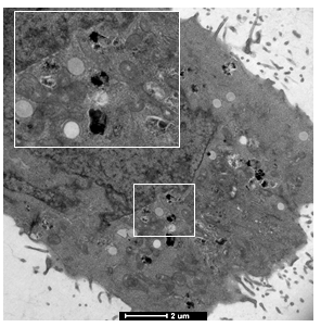
Image credit: Emily Guggenheim, UoB |
Particle localisation in organisms
- Tissue needs to be embedded in epoxy resin and dehydrated (in ethanol) to preserve the structure. Once dried, the sample can be cut into thin (100 micron) slices using an ultramicrotome.
- If needed, the sample can be stained to enhance the contrast or highlight specific organelles.
The image shown here is of iron oxide nanoparticles localised in A559 cells, with a slice thickness of 150 microns.
See NanoFASE procedure TEM-tissue localisation.
|
Contact
 Xianjin Cui
University of Birmingham (UoB)
 Iseult Lynch
University of Birmingham (UoB)
Email: i.lynch@bham.ac.uk